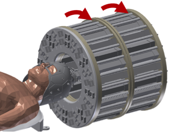
The cost and strict siting requirement of conventional high-field MRI scanners prevent the use of MRI outside of hospital radiology suites and necessitates that patients be transported to the scanner suite, precluding point-of-care imaging. To address this, we introduced a low-cost portable MRI scanner using a novel architecture [1] that could facilitate imaging in untraditional sites, like battlefield hospitals, senior centers, rural clinics, ambulances or a patient’s bedside. To enable this, we designed a lightweight, head-only, permanent magnet (Fig. 1).
The magnet size and shape were greatly reduced by allowing the field be low-field (~ 0.1T) and inhomogeneous (~1000x less homogeneity than typical MRI magnets). We note that allowing the magnet to be field not only reduces the size, but is also needed to ensure safety in unconventional locations. Instead of viewing the magnet inhomogeniety as a nuisance, we leverage it as the MRI spatial encoding magnetic (SEM) field, replacing the power-consuming, acoustically loud electromagnetic gradient coil hardware conventionally employed. This built-in encoding field is modulated by mechanical rotation of the magnet, while generalized projections of the object onto the SEM are acquired in the form of a spin-echo train. To reconstruct images, a general forward model approach is taken, in which an encoding matrix is calculated that relates the image to the data.
Transmit Array Spatial Encoding (TRASE) [2] was incorporated for encoding along the Halbach cylinder axis using broadband, frequency-swept WURST RF pulses [3]. This enabled preliminary 3D imaging in the prototype scanner [4]. Unlike the prototype Halbach magnet, the adult-size magnet for human brain imaging was designed with a tailored SEM [5]. The human-scale magnet is a sparse Halbach cylinder magnet made up of an optimized arrangement of NdFeB cube to create a desirable encoding field shape. This 122kg magnet design allows for higher resolution uniformity compared to the first prototype magnet, which is demonstrated in the 1cm lemon-slice image in Fig. 2. The complete scanner has been assembled on a cart which can easily be transported to a patient bedside and operates from a standard power outlet (Fig. 3).
[1] C. Z. Cooley et al., “Two-dimensional imaging in a lightweight portable MRI scanner without gradient coils,” Magn. Reson. Med., vol. 73, no. 2, pp. 872–883, Feb. 2015.
[2] J. C. Sharp and S. B. King, “MRI using radiofrequency magnetic field phase gradients,” Magn Reson Med, vol. 63, no. 1, pp. 151–161, 2010.
[3] J. P. Stockmann, C. Z. Cooley, B. Guerin, M. S. Rosen, and L. L. Wald, “Transmit Array Spatial Encoding (TRASE) using broadband WURST pulses for RF spatial encoding in inhomogeneous B0 fields,” J. Magn. Reson., vol. 268, pp. 36–48, Jul. 2016.
[4] C. Z. Cooley, J. P. Stockmann, M. Sarracanie, M. S. Rosen, and L. L. Wald, “3D Imaging in a Portable MRI Scanner Using Rotating Spatial Encoding Magnetic Fields and Transmit Array Spatial Encoding (TRASE),” Proc. 23rd Annu. Meet. ISMRM Tor. 2015, p. 0703, 2015.
[5] C. Z. Cooley et al., “Improved Uniformity of the Spatial PSF for Portable MRI Using an Optimized Rotating Magnet,” Proc. 25th Annu. Meet. ISMRM Honol. 2017, p. 1049, 2017.