Our group plays a leading role in the clinical validation and translation of new MRI methods, including efficient parallel imaging and reconstruction methods, motion correction and artifact reduction strategies, and machine learning assisted MRI reconstruction.
Fast 3D Imaging with Wave-CAIPI
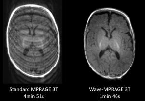
Wave-CAIPI provides highly efficient parallel imaging by taking maximal advantage of the 3D spatial information provided by multi-channel receiver coil sensitivity profiles. Through an active collaboration with the MGH main campus, we are leading the translation and validation of this technique for a variety of clinical applications including evaluation of hemorrhage using susceptibility-weighted imaging (SWI), evaluation of brain tissue volumes using T1-weighted MPRAGE, and evaluation of white matter lesions and cortical pathology using 3D SPACE FLAIR.
Ultra-Fast 2D Imaging with Multi-Shot Echoplanar Imaging (MS-EPI)
Echo-planar imaging is highly efficient, but diagnostic image quality is limited by factors such as poor tissue contrast, artifacts such as geometric distortion and signal dropout, and image noise. We have developed methods to improve image quality in EPI acquisitions to facilitate clinical adoption of these highly efficient techniques, including contrast preparation to take advantage of magnetization transfer effects, optimization of interleaved MS-EPI readouts, incorporation of simultaneous multislice (SMS) acquisition, and use of machine learning assisted reconstruction to provide “intelligent denoising” at high acceleration factors. This combination of approaches yields image quality that approaches conventional turbo spin-echo based images in a fraction of the acquisition time.
Selected References
- Bilgic V, Gagoski BA, Cauley SF, Fan AP, Polimeni JR, Grant PE, Wald LL, Setsompop K. Wave-CAIPI for highly accelerated 3D imaging. Magnetic Resonance in Medicine. 2015; 73(6): 2152–2162.
- Polak D, Cauley S, Huang SY, Longo MG, Conklin J, Bilgic B, Ohringer N, Raithel E, Bachert P, Wald LL, Setsompop K. A highly-accelerated volumetric brain examination using optimized Wave-CAIPI encoding. Journal of Magnetic Resonance Imaging. 2019; 50(3):961-974.
- Conklin J, Longo MGF, Cauley SF, Setsompop K, González RG, Schaefer PW, Kirsch JE, Rapalino O, Huang SY, Validation of highly-accelerated wave-CAIPI SWI compared to conventional SWI and T2*-weighted gradient-echo for routine clinical brain MRI at 3T. American Journal of Neuroradiology. 2019; 40(12):2073-2080.
- Longo MGF, Conklin J, Cauley SF, Setsompop K, Tian Q, Polak D, Splitthoff D, Liu W, González RG, Schaefer PW, Kirsch JE, Rapalino O, Huang SY. Evaluation of ultrafast wave-CAIPI magnetization prepared-rapid gradient-echo (MPRAGE) for visual grading and automated measurement of brain tissue volume. American Journal of Neuroradiology. 2020; 41(8):1388-1396.
- Filho ALG, Conklin J, Longo MGF, Cauley SF, Polak D, Liu W, Splitthoff DN, Lo W, Kirsch JE, Setsompop K, Schaefer PW, Huang SY, Rapalino O. Accelerated post-contrast Wave-CAIPI T1 SPACE achieves equivalent diagnostic performance compared with Standard T1 SPACE for the detection of brain metastases in clinical 3T MRI. Frontiers in Neurology. 2020 (Accepted).
- Lo W, Setsompop K, Liao C, Huang SY, Conklin J, Cauley S, Liu W, Clifford B, Bollmann S, Cao X, Zhang Z, Polak D, Splitthoff N, Feiweier T, Tian Q, Cho J, Kirsch J, Giri S, Rapalino O, Schaefer PW, Wald LL, Bilgic B. A comprehensive distortion-free 2-minute brain MR examination using BUDA and wave-CAIPI. International Society for Magnetic Resonance in Medicine, 2020. On-line virtual meeting.
- Conklin J, Clifford B, Bollmann S, Lo WC, Bilgic B, Cauley S, Setsompop K, Feiweier T, Kirsch J, Gonzalez RG, Schaefer P, Rapalino O, Huang SY. Comprehensive multi-shot EPI protocol for high-quality clinical brain imaging in 3 minutes. International Society for Magnetic Resonance in Medicine, 2020. On-line virtual meeting.